


Electron Microscopy
The imaging team provides a wide range of tools, training, and expertise, with a primary focus on providing access to world-class advanced electron microscopy instrumentation to the CNS community.
Electron Microscopy
The electron microscopy facilities at the Center for Nanoscale Systems (CNS) have been designed to enable cutting edge imaging and analysis research across the physical and biological sciences. The electron microscopy suites were custom-designed to house our sensitive microscopes, with minimal perturbations to imaging from environmental and other external factors. These facilities are supported by dedicated technical staff, with expertise in many aspects of electron microscopy and related techniques.
Several high resolution electron microscopes are available, including aberration corrected instruments, specialized cryo-electron microscopes and advanced analytical detectors. We have focused ion beam tools, which can be used for sample preparation, patterning, milling and in-situ analysis. Our high resolution scanning electron microscopes are equipped with a variety of detectors and are used to study a broad range of specimens. Dedicated spaces, fully equipped with tools for preparing samples, from hard materials and biological specimens, are also provided.
libra_200_02_1
sem_evo_02_1
2016-11-17 14.22.21-cryosem_t.baccata_leaf1
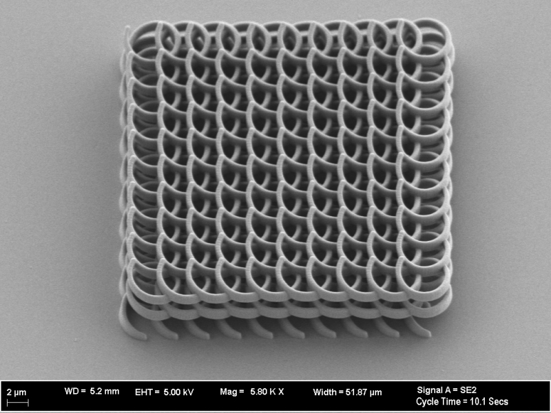
2016-12-14 15.27.16-nanoscribe_2_27_0

2017-1-17 11.0.32-nb_edited2
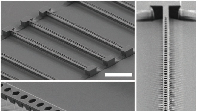
2016-12-14 15.27.16-loncar_03
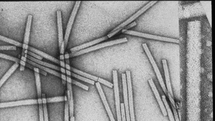
tmv
cds
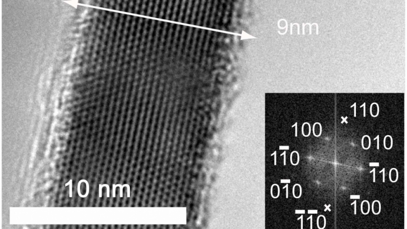
inasnanowire
hiv_trimer_lightup
proteasome raw image
block copolymer
160604 H206 AlGaN final_001
Finished Lamella SE Austin Cedric Ag70Au30

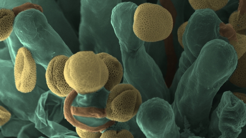
arabidopsis-ovules
0v_esb115
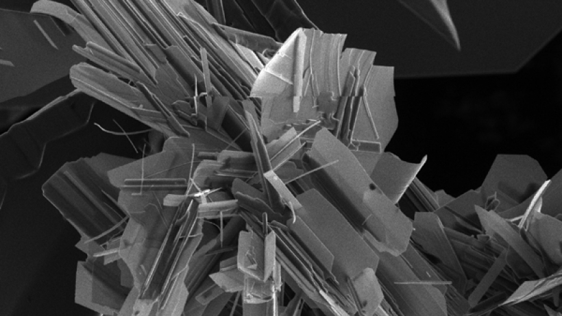
figure3
arabidopsis-stigma
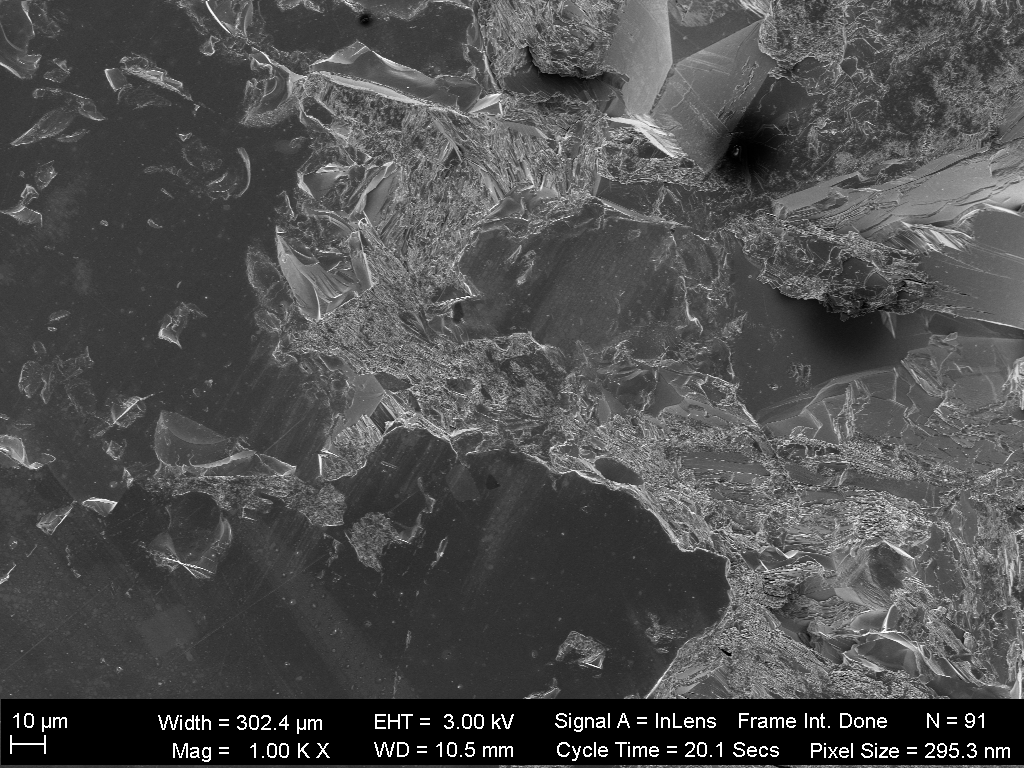
emery Inlens 005
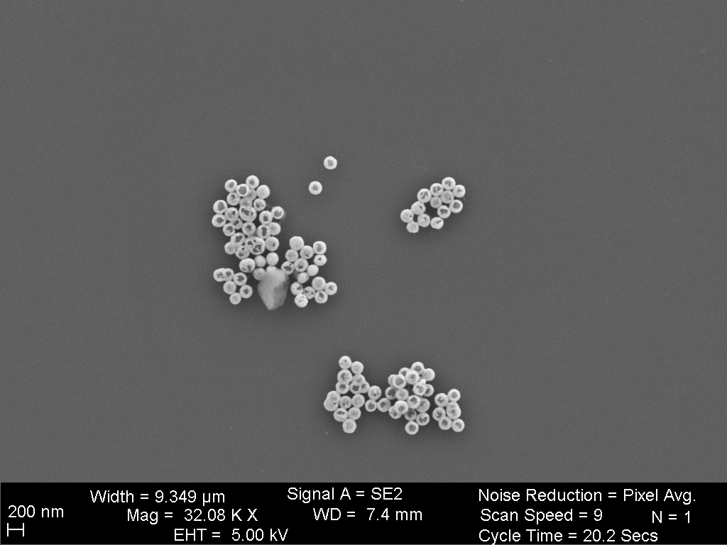
beads_00
Transmission Electron Microscopy TEM
Transmission Electron Microscopy is one of our core competencies at the Center for Nanoscale Systems. CNS is home to a state-of-the-art collection of high-resolution, aberration-corrected, and cryogenic TEMs. These allow the CNS community, with training and guidance from our knowledgeable staff members, to visualize and analyze their specimens at ultra-high spatial resolution.
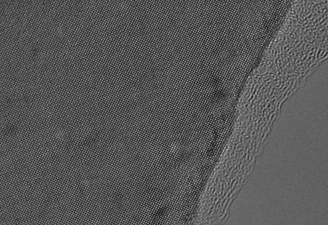
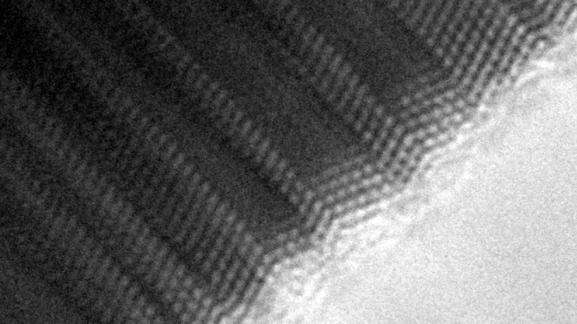
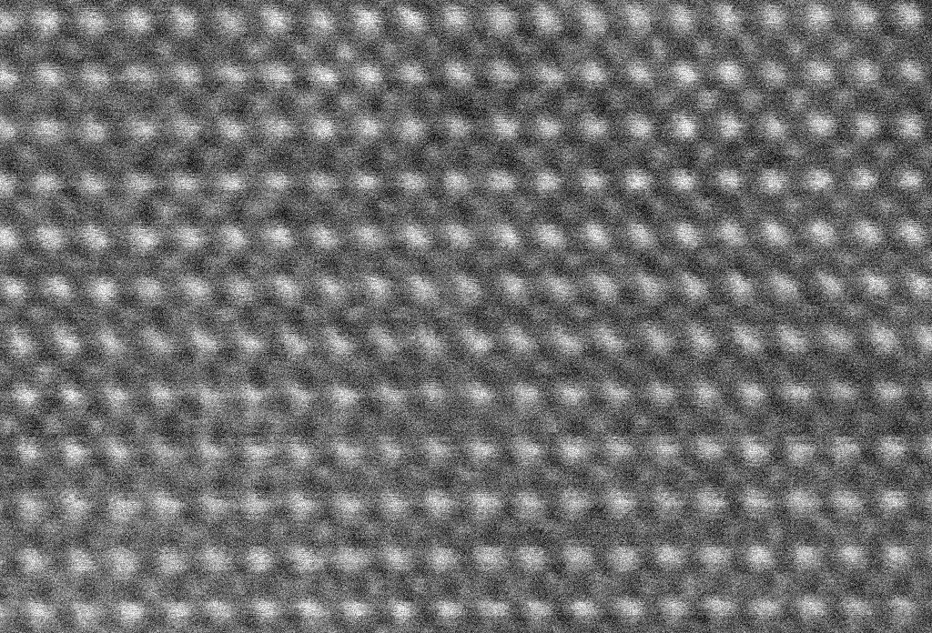
High Resolution TEM
High resolution transmission electron microscopy (HRTEM) is a powerful tool for materials analysis. This technique provides visualization of the atomic lattice, providing important information about a materials structure, defects, homogeneity and its composition.
At CNS, we have four HRTEMs available for materials analysis. Each HRTEM is capable of high resolution imaging and diffraction analysis. A dedicated diffraction camera, EDS and/or EELS detectors are available on some instruments. We have a selection of specialized specimen holders available for advanced TEM imaging and analysis.
In-situ electron microscopy allows for real-time visualization and analysis of chemical reactions and other dynamic processes. We have several customized holders available for in-situ TEM/STEM experiments.
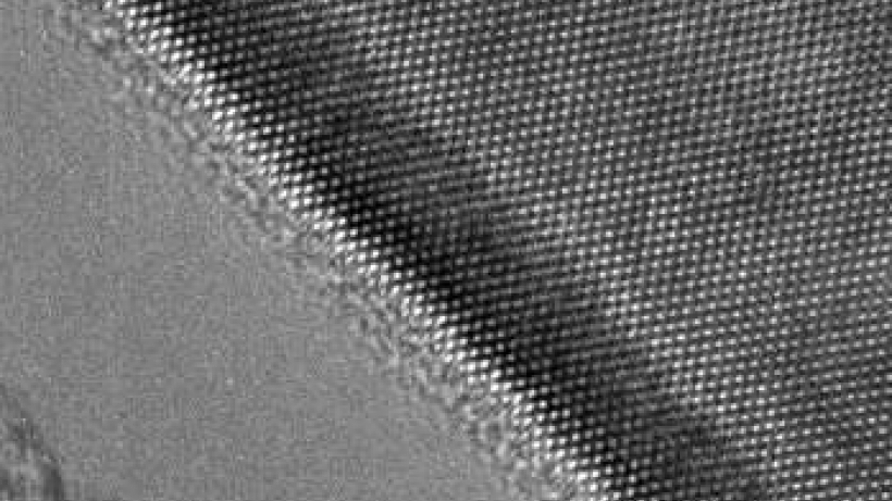
Scanning Transmission Electron Microscopy
The technique of Scanning Transmission Electron Microscopy (STEM) is often complimentary to TEM imaging. In this approach, a focused beam of electrons is rastered across the specimen sample, and the transmitted beam is analyzed. Typical STEM analyses include dark field imaging, bright field imaging, and spatially resolved analytical signals such as energy dispersive x-ray spectroscopy (EDS) or electron energy loss spectroscopy (EELS).
The Center for Nanoscale Systems has two STEM instruments, which enable high resolution imaging combined with analytical analysis. One of these instruments is a probe corrected cold FEG system that offers very high spatial and spectral resolution, combined with multiple imaging and analytical detectors.
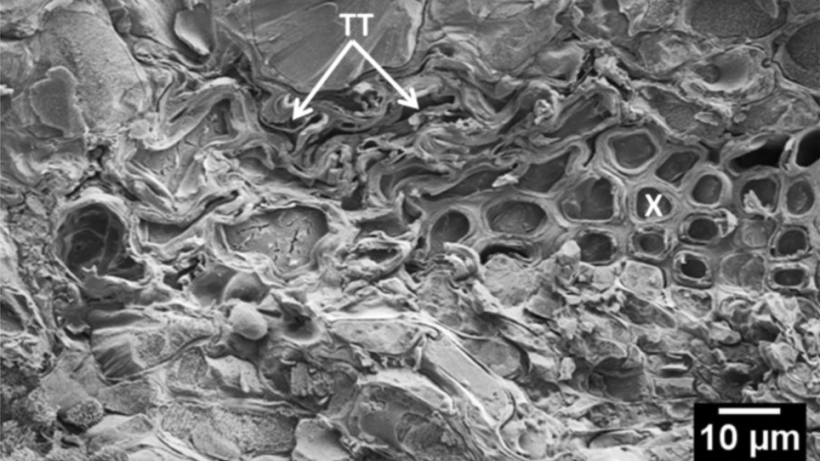
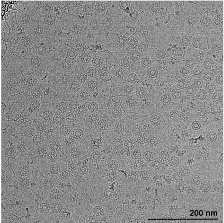
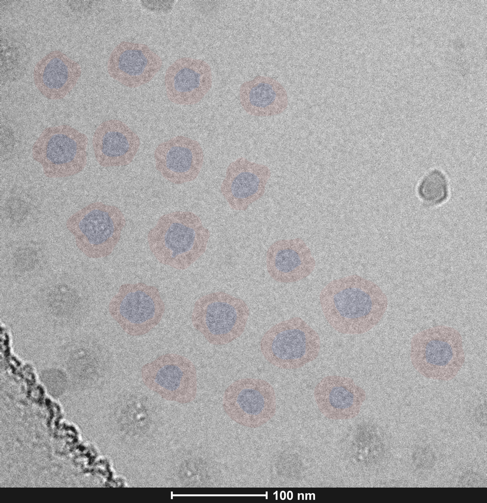
Biological Electron Microscopy
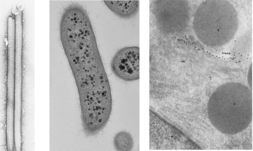
When preparing biological specimens for transmission electron microscopy , every step of the procedure affects the quality of the final images. Therefore, it is critical to process material according to prescribed protocols and understand what is happening in each step.
At CNS users can be trained on these sample preparation methods for a variety or sample types.
Negative staining is an established method for imaging the structure of proteins, single particles and small organisms, by contrasting a thin specimen with an optically opaque fluid. Routine morphology allows for specially prepared samples to be sectioned and high resolution of cellular infrastructure can be examined. Immuno cyto chemistry allows for the localization of proteins with in mildly fixed material. Users can be trained at CNS in these methodologies, or request service, or a combination of service and training depending on your research needs.
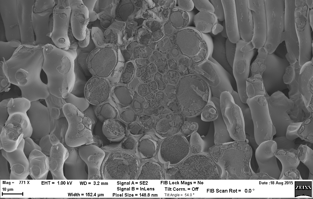
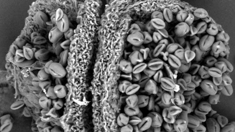
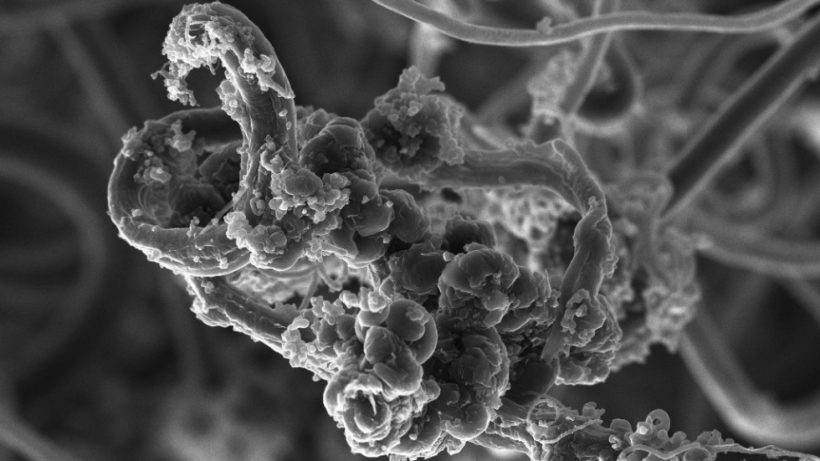
Cryo-SEM
The technique of cryo-SEM is a powerful method to study bulk hydrated specimens. These samples are not readily suitable for conventional SEM analysis due to their incompatibility with vacuum conditions. In cryo-SEM, the specimen is rapidly frozen and then imaged at cryogenic temperatures; this allows the integrity of the specimen’s structure to be maintained for imaging without using processing techniques such as dehydration or chemical fixation.
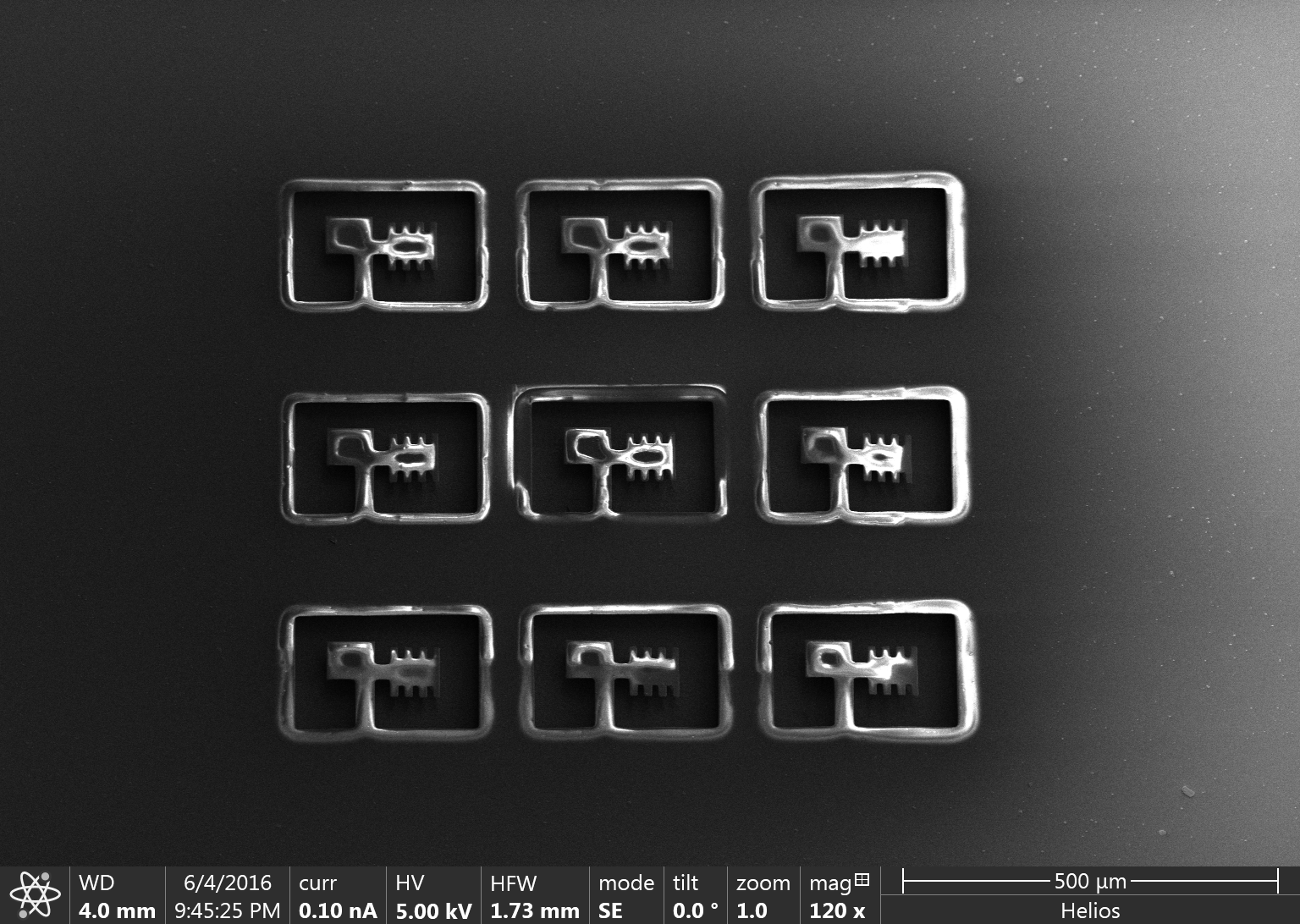
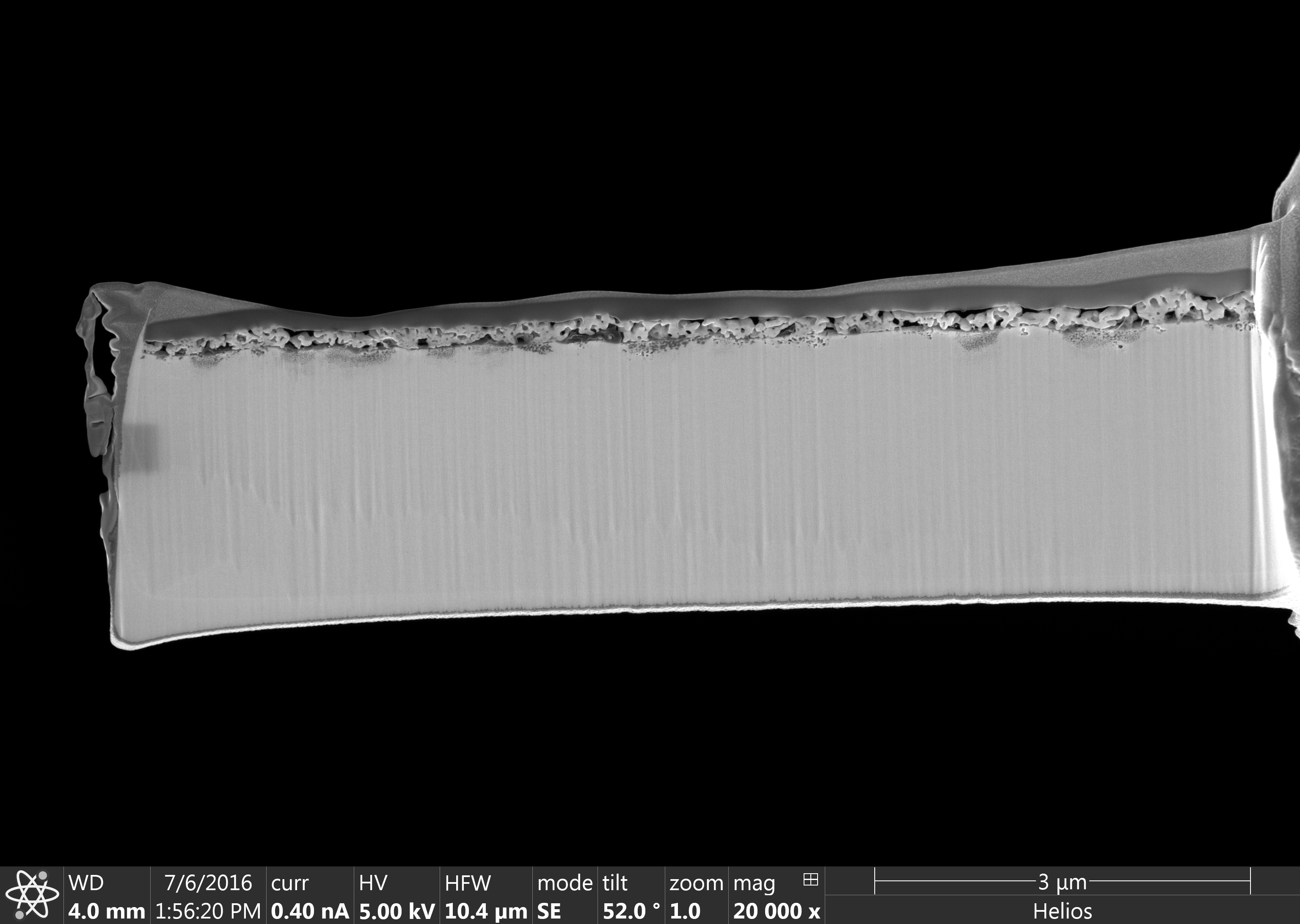
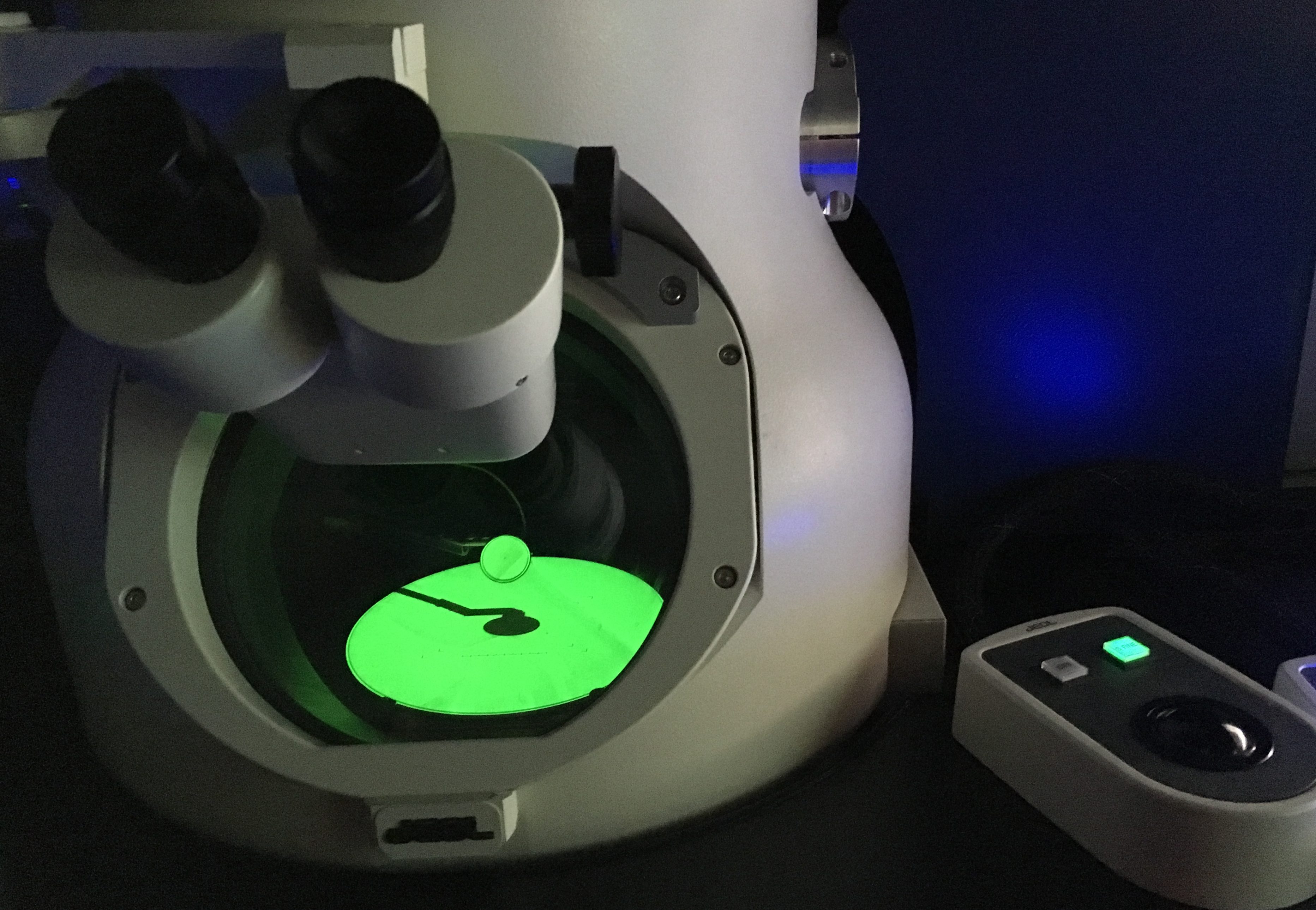
Focused Ion Beam
Focused Ion Beam systems use a finely focused beam of gallium ions to remove nanometers of material or deposit metals and insulators using gaseous precursors. These tools were originally developed for the semiconductor industry but have been readily implemented in the biological sciences, material sciences, applied physics, and geology fields.
At the CNS, we offer two dual beam FIB-SEM systems. These instruments have advanced milling, deposition, imaging and analytical capabilities. Our FIBs are particularly adept at preparing thin TEM specimens, high precision atom probe tips, and performing three dimensional imaging/analysis via the slice-and-view technique.
Scanning Electron Microscopy
Scanning electron microscopy (SEM) is a versatile tool for studying surface topography from the nanoscale to macroscale. In addition, the local surface chemical composition and crystallography can be studies using SEM-based techniques such as energy dispersive x-ray spectroscopy (EDS), wavelength dispersive spectroscopy (WDS) and electron backscatter diffraction (EBSD).
At the CNS, we have three high resolution SEM systems, each with a different combination of detectors.